New device for near-infrared fluorescence imaging in open surgery
The technique of near infrared (NIR) fluorescence imaging with the use of indocyanine green (ICG) is gaining more and more interest and recognition among surgeons of various specialties. It is used for:
- assessment of tissue perfusion of anastomoses in microsurgery (free flaps, stitched flaps), used in the assessment of wound demarcation or post-infarction injuries (visualisation of necrosis)
- evaluation of limb amputation stumps
- visualisation of lymphatic pathways (lymphography, ARM) – eligibility for lymphatic reconstruction (LVA, LNT),
- assessment of tissue perfusion during anastomosis in colorectal surgery,
- Mapping of tumour boundaries (secondary liver metastases, HCC)
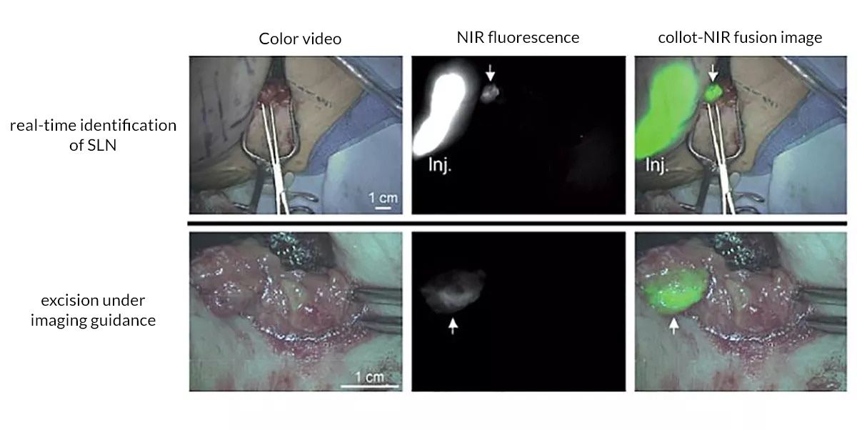
- localization of the sentinel node (SLN) during tumour staging in general surgery, urology and gynaecology.
- Detection of parathyroid glands to avoid damage during thyroidectomy; etc.
An innovative solution for the diagnosis of lymphoedema
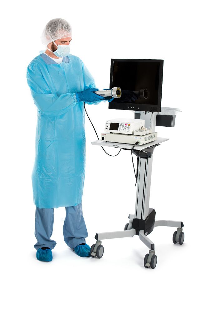
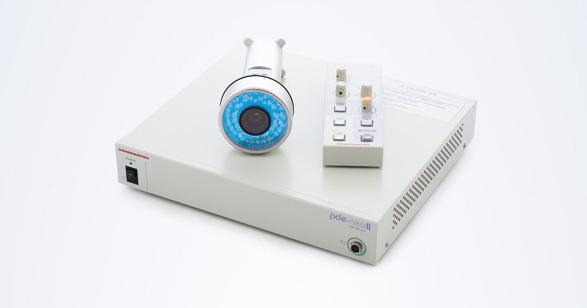
This is a revolutionary indocyanine green fluorescence imaging device. PDE-Neo is used for visualisation of blood flow during surgery. Applications include assessment of tissue perfusion and associated free lobe circulation.
PDE Neo II can also be used for visualisation of lymphatic pathways in super reconstructive microsurgery. This innovative technique holds great promise for mapping patients with lymphoedema using ICG fluorescence.
The device, which offers the highest sensitivity on the market today, enables real-time visualisation of lymph flow, qualitative analysis of the images obtained and comparison of, for example, the status of dilated or damaged vessels over the course of consecutive days.
The device has a fluorescence mapping function with ultra-high visibility of the ICG dye and visualisation of lymphatic vessels in green. This makes it possible to precisely map the superficial lymphatic system in the patient’s tissues.
Three ICG visualisation modes:
- White light used for analysis of anatomical aspects and focus calibration;
- Fluorescence for detection of ICG – indocyanine green
- Fluorescence mapping to display digitally enhanced contrast between ICG and surrounding area
Features:
- The fluorescence mapping feature creates a high contrast image by applying green contrast to near infrared fluorescence images. Through a unique digital subtraction process, the non-fluorescent image background can be independently adjusted to the surgeon’s preference.
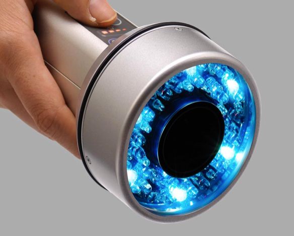
- Focus adjustment (near-far)
By rotating the focus knob on the camera, focused images can be observed at different distances, depending on the working distance. - Colour and B/W image
Easily switch between black-and-white and colour image.
A useful option when comparing anatomy with a fluorescence image. - White LED light
The LED white light option illuminates the operating field without losing the fluorescence image. This is particularly useful when the lights in the OR have been turned off to avoid interference with the fluorescent image. - Status display
Enabling the status option will display the pde-neo II settings in real time. It allows the user to quickly check the current brightness, contrast and excitation light settings.
Advantages:
- Enhanced fluorescence visibility
- Built-in full colour / white light camera
- Tool for colour mapping and fluorescence analysis
- Works with ICG fluorescence
- Compact handheld design
- Manual calibration possible for operator and separate console for assistant